We use cookies on this website to enhance your user experience. Please read our cookie policy for additional information and a complete overview. We will only use cookies if you consent by clicking on “Accept all cookies”. You can always manage your preferences via the settings of your browser. Please note that certain media will only be available if you have accepted the applicable cookies.
PreOp changes to Persyst ESI supported by Epilog
Please go to: www.persyst.com/persystESI
We would like to assure you that in the meantime nothing changes for you, the PreOp solution is continuing as normal. Of course, if you are interested in getting a demo and testing the new solution for free, please let us know.
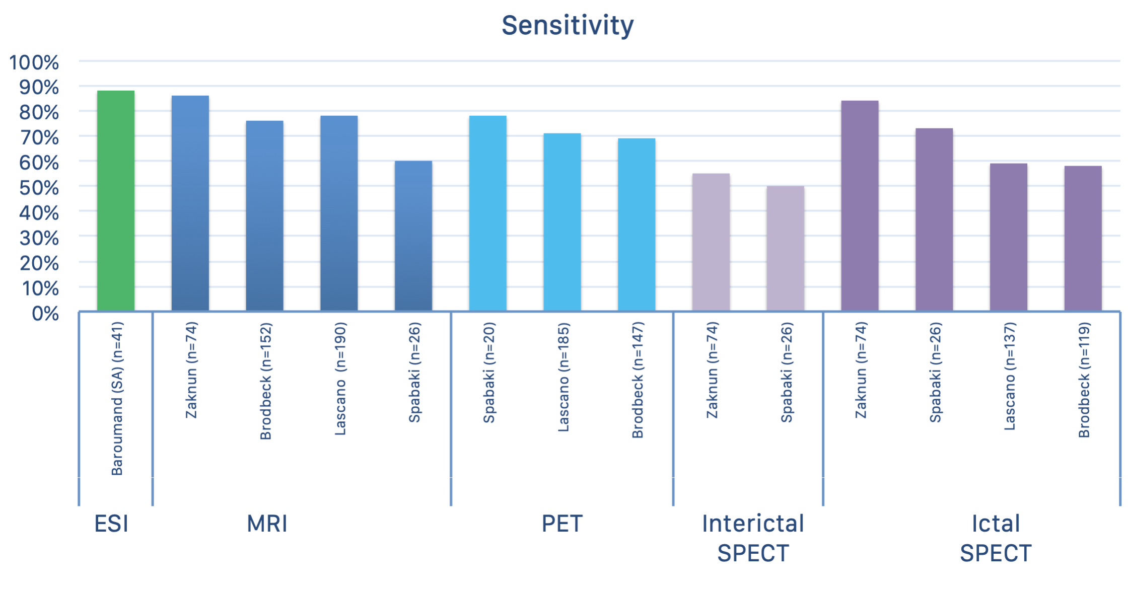
Electrical source imaging can provide valuable insights in the preoperative evaluation for refractory epilepsy. However, such advanced EEG analysis is a time-consuming and labour-intensive process. Epilog PreOp offers an efficient and clinically validated alternative with a sensitivity of 88% after interpretation by an expert.
Features
Epilog PreOp uses accurate and clinically validated techniques for spike detection and clustering.
A patient-specific head model that includes six tissue types (scalp, skull, CSF, air cavities, gray and white matter) is generated from your patient’s MRI. The distrinction between gray and white matter and modeling is crucial for the most accurate localization of epileptic activity in 3D within the brain. This is not implemented in other software packages.
The results of the analysis are provided in a report that contains all relevant information. The reconstructed epileptic activity can also be visualized using our 3D viewer, or exported to the PACS system of your hospital.
CLINICALLY VALIDATED
The complete Epilog PreOp processing pipeline was validated on typical clinical long-term EEG recordings (25 electrodes). The results are depicted below and show that a semi-automated interpretation of electrical source imaging generated by our pipeline obtains higher sensitivity than MRI, SPECT and PET during the preoperative evaluation.
*Baroumand, Amir G., et al. “Automated EEG source imaging: A retrospective, blinded clinical validation study.” Clinical Neurophysiology 129.11 (2018): 2403-2410.

