We use cookies on this website to enhance your user experience. Please read our cookie policy for additional information and a complete overview. We will only use cookies if you consent by clicking on “Accept all cookies”. You can always manage your preferences via the settings of your browser. Please note that certain media will only be available if you have accepted the applicable cookies.

Source localization of epileptic seizures
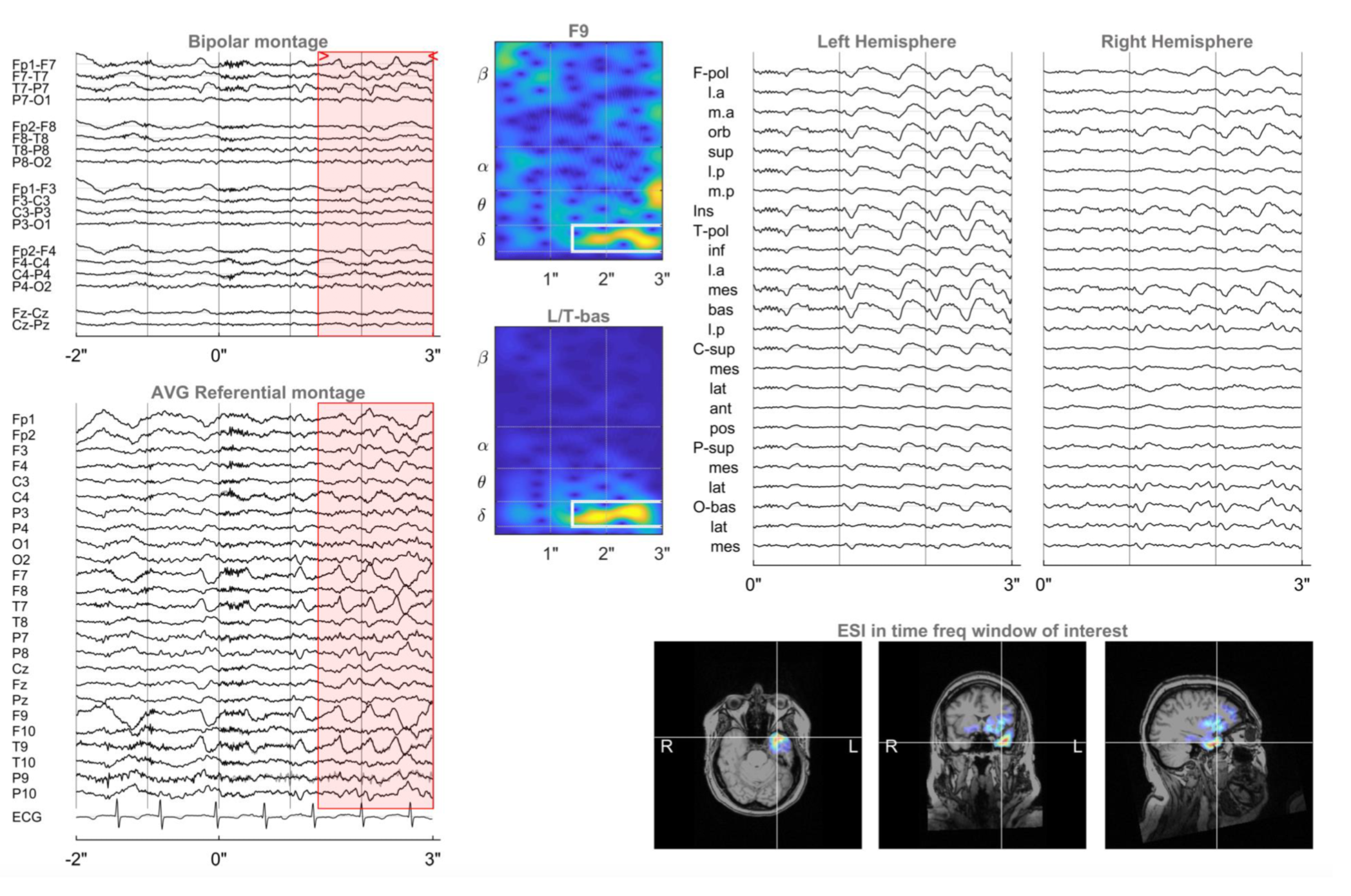
The ictal ESI technology is based on 25 years of research at Ghent University, Ghent University Hospital and Epilog. The technology is validated by the top experts in the field resulting in a 74% accuracy.
Features

Manual onset annotation
Mark the electrographic onset of the seizures in the EEG of your patient.

3D patient-specific source localization
A patient-specific head model that includes six tissue types (scalp, skull, CSF, air cavities, gray and white matter) is generated from your patient’s MRI. The distrinction between gray and white matter and modeling is crucial. >15000 dipoles are modeled for accurate sublobar localization.

Concise online report and 3D-viewer
The results of the analysis are provided in a report that contains all relevant information. The reconstructed epileptic activity can also be visualized using our 3D viewer, or exported to the PACS system of your hospital.